Express Grants Tackle 3D Scanning Technology
Team Investigates Possibilities for Future Uses in the Classroom
When NC State faculty find themselves looking to solve pedagogical challenges, they often turn to DELTA for solutions. Specifically, DELTA Express Grants offer help to explore specific innovative teaching methods or technologies. These grants usually involve focused but limited DELTA support, typically over the course of a single semester.
A DELTA team recently shared the outcomes from two DELTA Express Grants for the following courses within the Department of Entomology and Plant Pathology that had overlapping objectives that included 3D object scanning:
Both grant proposals sought 3D scans of available specimens to improve student access for further study and fit well with the Express Grant format. Additionally, these grants aligned with DELTA’s goals of investigating methods to expand content capture services and to leverage the use of 3D objects in the classroom with available media formats.
3D object scanning, or photogrammetry, is a process that uses photographs or videos of real-world objects or environments to create digital 3D models of those objects. It involves capturing many overlapping images of a structure, object or landscape.
According to Multimedia Specialist Stephen Waddell, the model created from photogrammetry can be a representation of the subject that then can be viewed from multiple angles and potentially used for animation or even interaction.
“These photos combined in some sort of photogrammetry software are ultimately used to produce the 3D model and reproduce the surface color,” Waddell explained.
Developing Partnerships
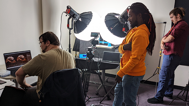
This exploration began a few years ago when the DELTA team worked with Principal Investigators Extension Associate Terri Billeisen and Alumni Association Distinguished Undergraduate Professor of Entomology Clyde Sorenson on the Express Grant for ENT 201 Insects and People. For this grant, the team assembled a macro photography rig for binding photo stacks at different angles to capture 3D objects typically too small for traditional photogrammetry practices.“For this work, we partnered with the Libraries to further enhance one of their studio spaces to support this effort throughout the duration of the project,” Waddell said. “We also worked directly with University Library Specialist Colin Keenan on organizing and structuring the photo shoots. He provided not just space but also new ideas, insights, views on the setup and a lot of equipment over the course of the photogrammetry effort.”
Meeting the Challenges
Although the team had a good starting point for the setup, they needed to make notable improvements to complete the photoshoot.
Director of Digital Media Innovation Michael Cuales clarified that macro photography and photogrammetry require precise camera settings and detailed specifications to provide the needed results. Thus stability, environment and light were critical factors to tackle these issues. Over the course of the photoshoot, the team modified the two access turntables and lighting used, providing significant improvements. “We leveraged expertise from Michael Castro and Relly Moorer from the Instructional Media Production (IMP) team, who brought an insane amount of light to the situation, which at one point began to melt our polarizing film,” Cuales said. “We quickly altered that set up!”
Getting the Desired Outcomes
The team began capturing images of small insects, such as crickets for the ENT 201: Insects and People course. Experimentation with different types of insects showed distortion or even large missing parts of the bug entirely. As the project went on, the outcomes of each photoshoot informed the next, and the team was able to better control the lighting, improve the optics and ultimately refine the results.
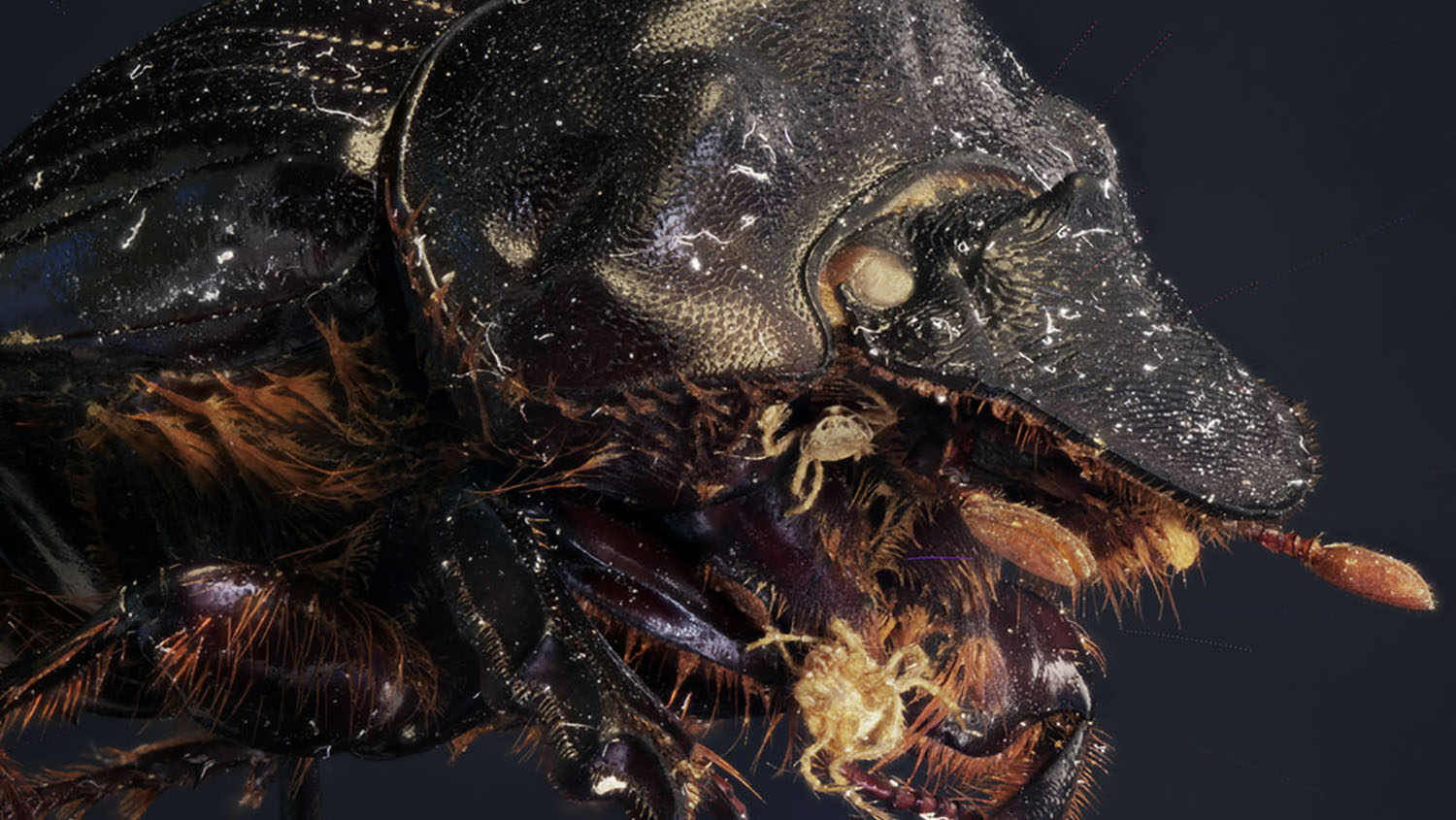
Settling on a beetle, the team achieved a reasonable scan to produce a 3D model. Previous experience with processing 3D scans made any clean up on the images fairly minimal to achieve an archival, 3D model with the added benefit of details undetectable by the human eye in the photos.
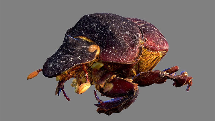
According to Waddell, the beetle was just one option in a range of possible choices for the project.
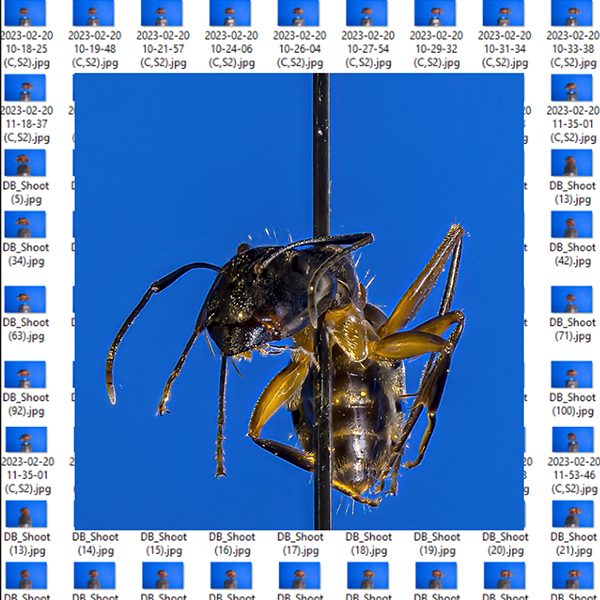
“The second 3D capture Express Grant that we worked with, ENT 132 Urban Pest Management, focused on an ant,” Associate Director Donnie Wrights said. “Although a hairy spider was considered, the ant provided enough challenges without the additional hair and legs.”
Wrights explained that the team remained focused on how to improve the overall experience for students and provide a resource they can come back to. “The desired outcomes for this project were to enable students to rotate and zoom in and out to examine specific structures,” Wrights said. “We want the students to closely examine the insects in order to understand how the insects improve, adapt and survive in their environment.”
Takeaways and Lessons Learned
For the ant imaging, the 3D model unfortunately never fully solidified, despite a number of iterations that moved closer to a successful outcome. However, each iteration did provide the team with ideas of the next steps that might be helpful when working with something on this scale and the challenges of making further improvements to the process.
“For ENT 132 Urban Pest Management, we were able to get hundreds of photos and create a high-resolution turnaround of the ant that students can use in the classroom,” Waddell said.
The team learned that one important factor to getting high-quality photos was having a space with a long process of stacking so that large runs of photos needed to proceed without interruption for up to nine or more hours. In addition, having more angles and elevations made it easier to avoid problems with single takes. They also realized that control of the lighting was still an issue, as some images were still showing glare and having issues with polarization.
“Getting a flat photo, both in lighting quality and geometry, is still difficult,” Waddell added.
NC State University Libraries is currently renovating two capture studios to expand services and media work. These studios will facilitate ongoing explorations and macro photogrammetry as well as new tests and technology, such as volumetric frame capture, markerless animation and further experiments into new media as they become available.
“3D scanning technology is rapidly evolving, making it easier to capture 3D representations of large areas, people and super small objects,” Cuales said. “We’ve invested in some known quantity methods for capture, like the use of drone-based photogrammetry. We’ve also invested in less conventional methods of scanning small objects, like an intra-oral scanner commonly used for dental work.”
The team that worked together to complete these projects and gather this information was large and included the following people:
- DELTA specialists (Broadcast and Emerging Media Specialist Michael Castro, Director Digital Media Innovation Michael Cuales, Broadcast and Emerging Media Specialist Relly Moorer, Multimedia Specialist Stephen Waddell, and Associate Director Donnie Wrights)
- Faculty Principal Investigators (Director and Training Coordinator Patricia Alder, Extension Associate Terri Billeisen, and Alumni Distinguished Undergraduate Professor of Entomology Clyde Sorenson)
- NC State University Libraries staff (Specialist Colin Keenan)
DELTA expects to expand its 3D scanning capacity and capabilities while investigating new ways to leverage the use of 3D objects in the classroom with existing media formats.
Want information about how to work together with DELTA to develop instructional technologies for your course? Learn more about the grant cycle and how to apply for a DELTA Express Grant.
- Categories: